
LUNGEIT KIT
Start lung EIT research with a complete setup - and keep the freedom to change it.
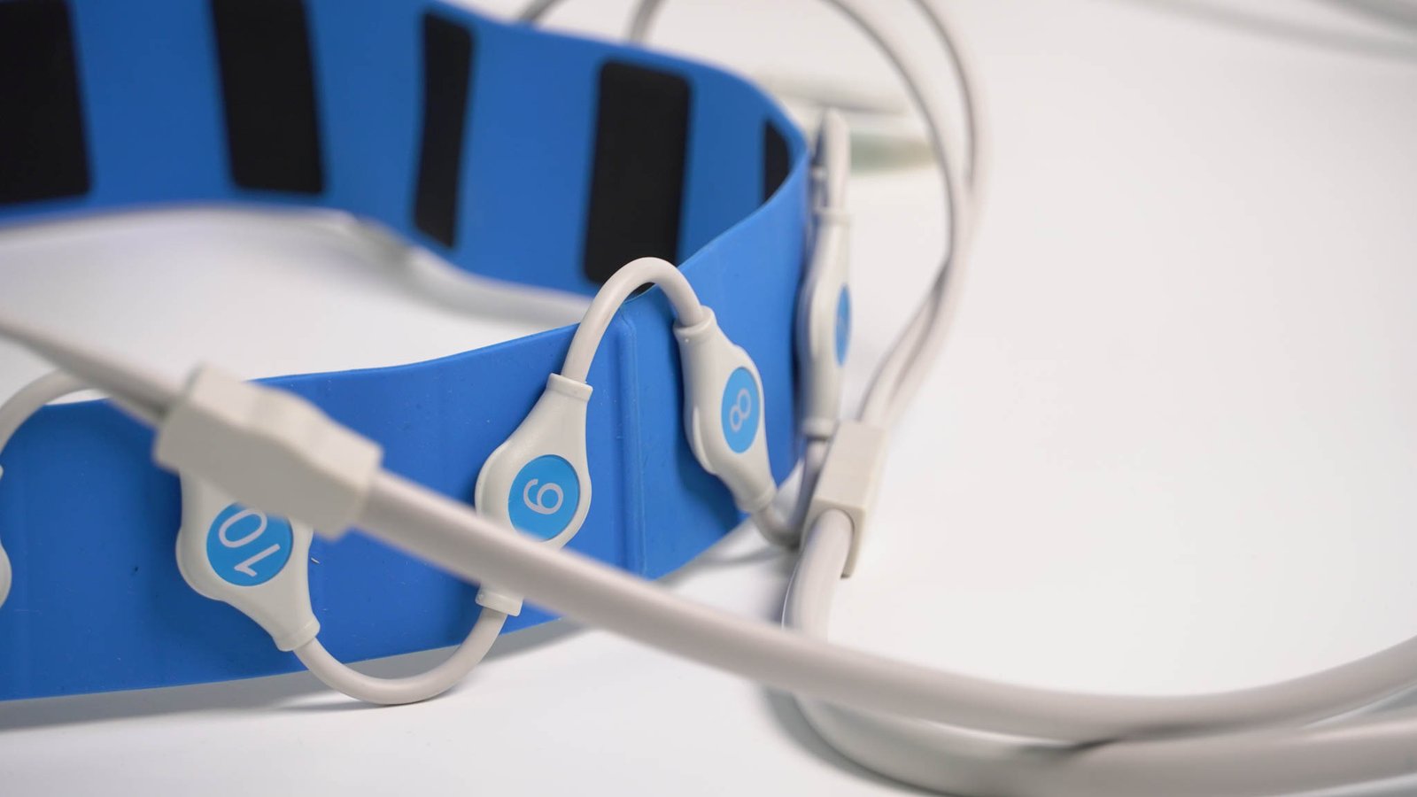
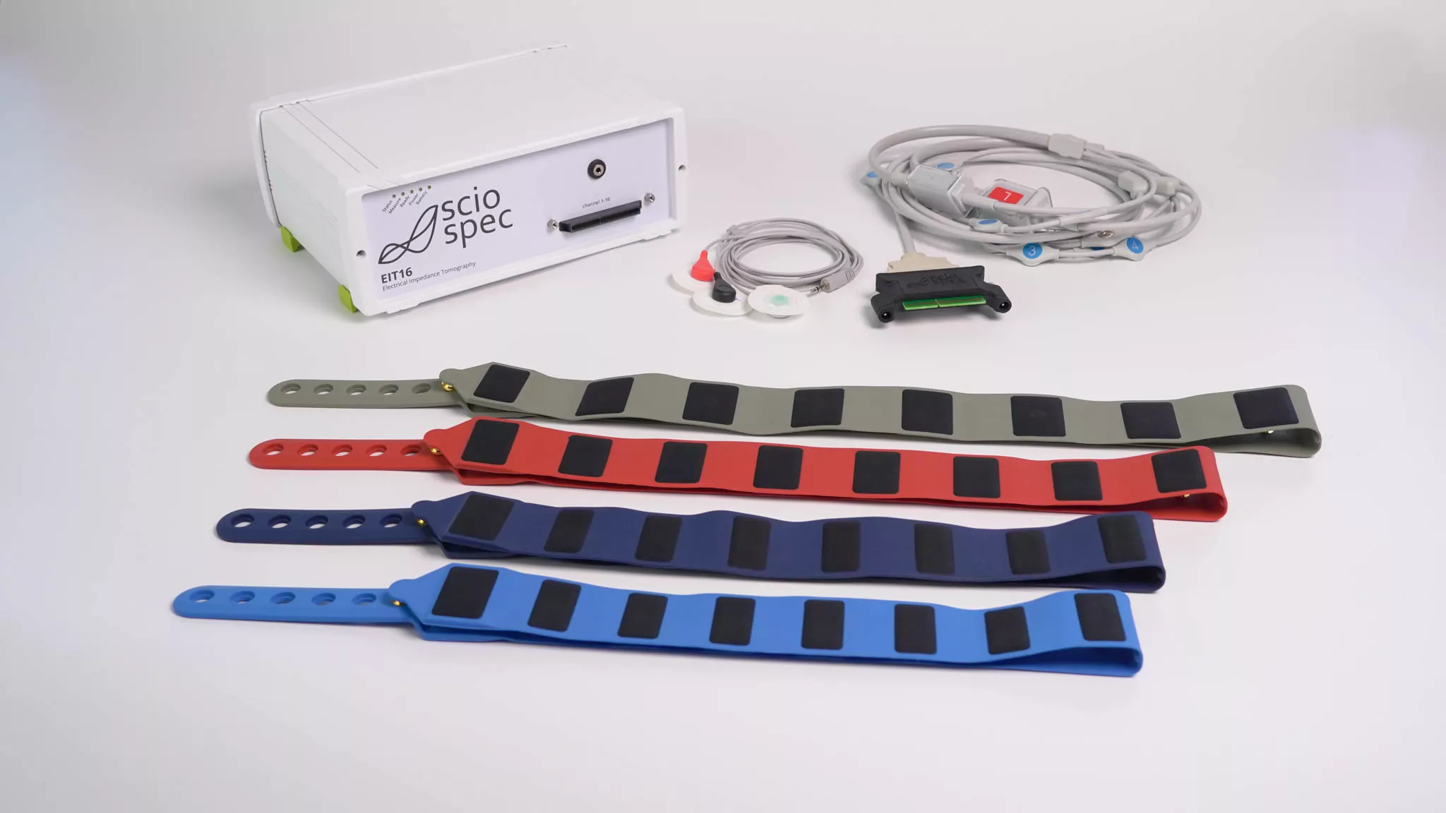
The LungEIT Kit brings together Sciospec’s dedicated 16-channel EIT system, 16-electrode lung belts, cabling, lung-imaging software and a safety-oriented configuration for human-connected research.
Instead of building the belt interface, isolation chain and basic imaging workflow yourself, you can start with a working pulmonary EIT setup.
That easy start does not turn the system into a closed clinical monitor. You can still change injection patterns and measurement settings, access raw EIT data, use your own reconstruction matrix, synchronize ECG or external equipment, and take the data into MATLAB/EIDORS.
- Complete 16-electrode lung EIT setup with belts in sizes S to XXL
- Dedicated EIT acquisition with frame rates up to 100 fps, configuration-dependent
- Lung imaging software plus raw-data, custom reconstruction and EIDORS-compatible workflows
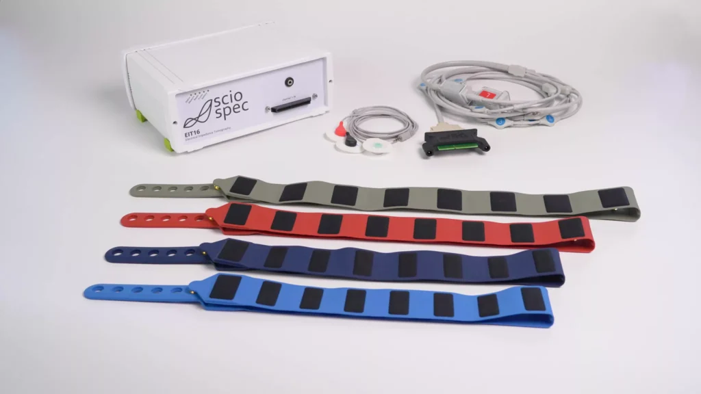
16 ELECTRODES
LUNG BELTS S-XXL
UP TO 100 FPS
DYNAMIC EIT
Capture breathing-related impedance changes with the dedicated EIT architecture; actual frame rate depends on the configured sequence and frequencies.
SAFETY-ORIENTED
RESEARCH CONFIGURATION
OPEN DATA
RECONSTRUCTION FREEDOM
Use the lung app, your own reconstruction matrix or raw EIT data in custom and EIDORS-based workflows.
From belt to lung image - without building the setup yourself.
For many pulmonary EIT projects, the time-consuming part is not the idea of EIT itself. It is getting the practical chain around the experiment to work: belts and cabling, acquisition, reconstruction, synchronization, data access and the electrical-safety concept for a human-connected setup.
The LungEIT Kit brings those pieces together around Sciospec’s dedicated EIT platform. Start with a working pulmonary workflow, then open it up as soon as the study needs different injection patterns, raw data, custom reconstruction or additional synchronized signals.
16-ELECTRODE LUNG BELT → CONFIGURE ACQUISITION → DYNAMIC EIT MEASUREMENT → LUNG IMAGE → RAW DATA / CUSTOM RECONSTRUCTION / EIDORS → OPTIONAL ECG & EXTERNAL SYNCHRONIZATION
Start with the lung study, not the instrument build
Use standard lung belts, defined cabling and the dedicated lung-imaging app instead of first developing the physical setup and basic imaging workflow yourself.
Change the measurement when the study asks for it
Adjust injection patterns, frequency, current and acquisition settings instead of being limited to one fixed monitoring protocol.
Keep access to the measurements behind the image
Start with Sciospec reconstruction, use your own reconstruction matrix or export raw frame data for custom processing and EIDORS/MATLAB workflows.
Synchronize EIT with ECG and external equipment
Available ECG synchronization and isolated I/O routes support studies that combine respiratory EIT with physiological signals or external equipment.
Technical Specifications
Key specifications for first technical qualification of the current 16-channel LungEIT Kit. Detailed EIT timing and electrical characteristics remain available in the dedicated EIT technical documentation.
Parameter
Specification
Details
LUNG EIT SETUP
Current standard channel count
16
The current product configurator identifies the 16-channel kit as the available standard LungEIT configuration
Lung electrode belts
16 electrodes
Standard belt sizes S, M, L, XL and XXL in current product material
EIT acquisition
Synchronous multichannel voltage acquisition
All measurement-channel voltages are sampled together for each active current-injection state
Injection sequence
Configurable
User-defined source/sink pairs and sequence within the EIT platform
MEASUREMENT
Frequency range
100 Hz - 1 MHz
Released dedicated-EIT specification
Frequency sweep
Up to 128 frequencies
Multifrequency EIT; more frequencies increase complete-frame time
Current excitation
100 nA - 10 mA peak
Released dedicated-EIT specification
Frame rate
0.1 - 100 fps
Configuration-dependent; determined by injection sequence, frequencies and acquisition settings
Signal extraction
Sciospec SineFit
Complex signal extraction in the dedicated EIT platform
SOFTWARE & DATA
Lung imaging software
Dedicated LungEIT application
Ready-to-use pulmonary EIT imaging workflow
Standard reconstruction
Time- and frequency-difference EIT
Sciospec reconstruction workflow
User-defined reconstruction
Custom reconstruction matrix
Use an application-specific matrix in the Sciospec workflow
Raw-data route
Raw EIT frame data
For custom processing and reconstruction
External analysis
MATLAB / EIDORS-compatible route
For research and custom algorithm workflows
SAFETY & INTEGRATION
Power
Medical-grade external power supply
Current LungEIT material specifies Insulation Class II / EN 60601-1-compliant power supply components
PC connection
Isolated full-speed USB
Instrument control and data transfer
Network connection
Ethernet + external medical-grade network isolator
Current material states the external isolator is used to provide the required isolation in the network path
Battery operation
Available in current LungEIT configuration
Supports operation without direct mains connection to the instrument
ECG
1-channel synchronized ECG
Available in current LungEIT product material; verify included/optional commercial status
External synchronization / I/O
IsoIO / hardware synchronization
For integration with external sensors and devices; verify current package status
AC coupling
External AC-coupling frontend
Application-specific route for blocking DC and providing galvanic isolation at the patient interface; verify current standard/optional status
ISX-3 EIT with 32/64-port array — Show the analyzer and multiplexer physically connected to a phantom, sensor or electrode structure so the product is immediately understood as more than a standard ISX-3.
Safety and integration hardware — power, isolation, battery and I/O components physically connected, with captions that state what is standard, optional or application-specific.
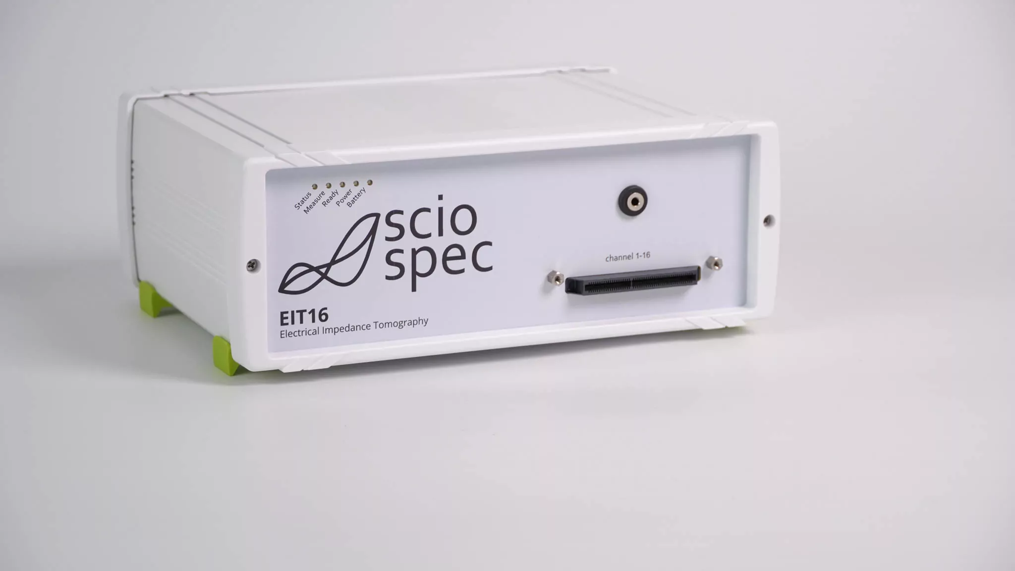
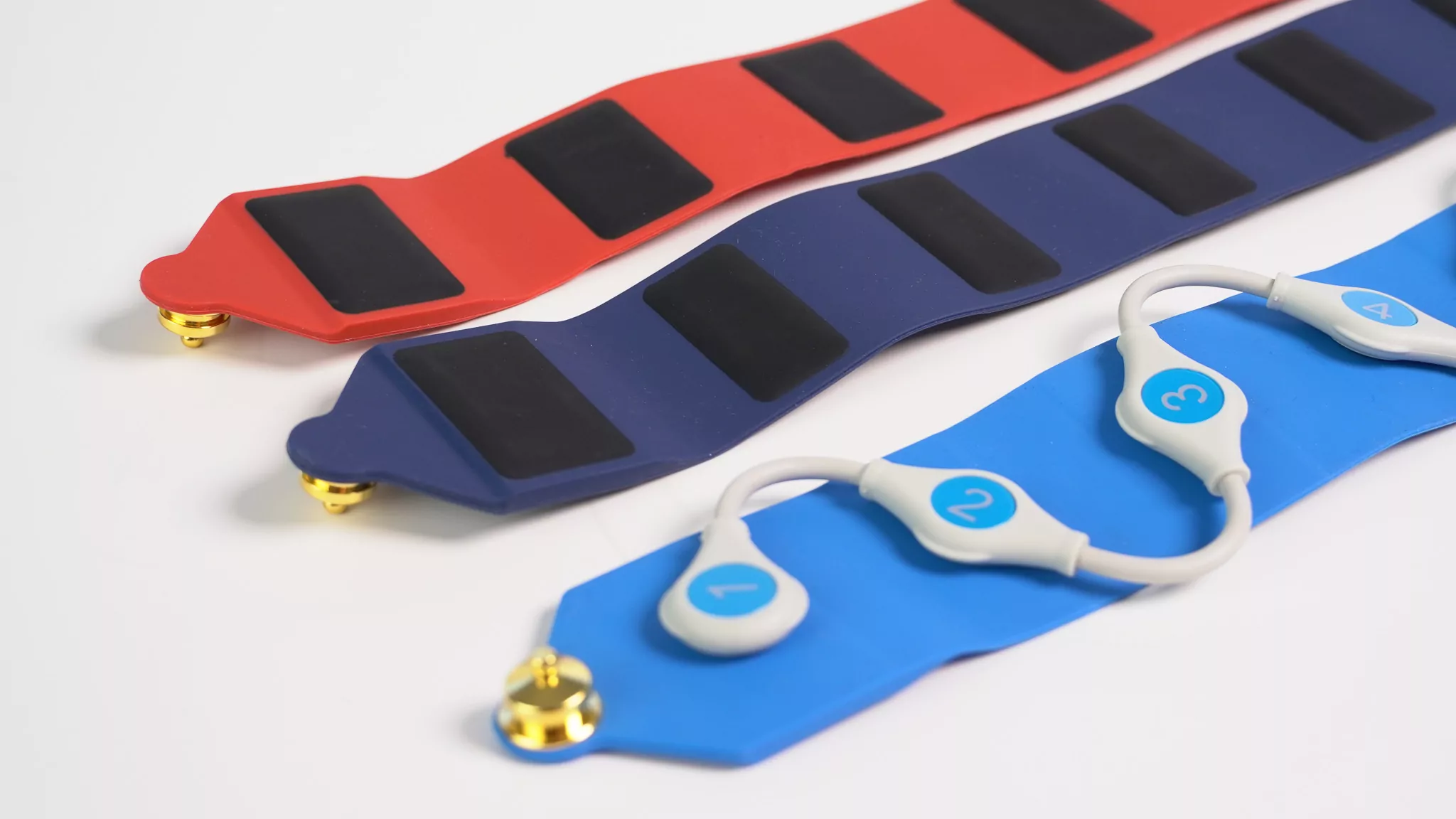
Made for clinical research — safe & easy, but with all the flexibility you need.
Visualize changes in regional ventilation
Use dynamic EIT to follow breathing-related impedance changes and compare ventilation distributions over time or between study conditions.
Study how ventilation changes with the protocol
Use repeated EIT measurements around respiratory interventions, positioning, rehabilitation or other controlled study conditions where regional change matters.
Work on the measurement underneath the image
Access raw data, define injection patterns and use your own reconstruction matrix or EIDORS-based workflow for method and algorithm development.
Develop new monitoring and diagnostic concepts
Use an open research platform to establish the measurement, belt/electrode concept, synchronization and analysis before separately engineering a dedicated medical or OEM system.
Safety features that make the step into clinical studies easier.
Medical-grade power and a protected enclosure
Use the medical-grade external power supply and safety-oriented enclosure modifications supplied for the LungEIT setup. Stand-alone battery operation is also available when the study benefits from operating without a direct mains connection to the instrument.
Isolated PC and network connections
Control and read out the system through the isolated full-speed USB interface. If Ethernet is used, the external medical-grade network isolator described in the LungEIT offer can be inserted into the communication path.
AC coupling when additional isolation is needed
An external AC-coupling frontend can block DC currents, limit low-frequency currents and galvanically isolate the instrument from the measurement object or patient interface when the study requires it.
Synchronize ECG and external research equipment
Use the IsoIO and hardware-synchronization routes to integrate external equipment, or add synchronized ECG when the study needs respiratory EIT and cardiac signals on the same timeline.
Configure the 16-channel LungEIT research setup
Loading configurator…
Get the lung image quickly. Keep access to the data behind it.
Get from measurement setup to a lung image quickly
Configure the measurement and view reconstructed lung EIT data in the dedicated application without first building your own imaging software.
Change the EIT setup when the study needs it
Adjust injection strategy and measurement settings when the study needs something beyond the standard workflow.
Use your own reconstruction matrix
Bring an application-specific reconstruction matrix into the workflow when the geometry or algorithm requires a different model.
Take the data into your own workflow
Export EIT frame data for custom processing, MATLAB/EIDORS-compatible reconstruction and your own analysis pipeline.
When should you choose the LungEIT Kit?
Choose it when routine bedside monitoring is the job
Established clinical systems are the natural choice when approved monitoring workflows, clinical metrics and routine bedside operation dominate. LungEIT is the research route when raw data, custom reconstruction, synchronized signals or application development matter.
Choose generic EIT when the lung-specific setup is unnecessary
EIT16 uses the same dedicated 16-channel measurement principle. Choose it when you need a general EIT research instrument rather than belts, lung imaging and a human-research-oriented system configuration.
Build the rig when the instrumentation itself is the research
A home-built system makes sense when current-source, acquisition, switching, safety architecture and software development are part of the scientific objective. LungEIT adds value when the pulmonary question is the real work.
Move beyond the standard kit when the electrode concept changes
The current standard LungEIT Kit is 16-channel. Research that needs more electrodes, several planes or a novel belt geometry should be reviewed against the EIT32/64/128+ platform or a custom development route.
Develop the measurement and study workflow before you freeze the device.
Establish the pulmonary measurement
Test belt/electrode concept, acquisition settings, reconstruction, synchronization and study workflow on the configurable research platform.
Stabilize the workflow
Define the belt sizes, interfaces, external signals, analysis and operating procedure required for repeatable studies.
Engineer the final system separately
When the requirements are mature, scope dedicated electronics, software, mechanics, compliance and manufacturing as a new development stage.
Frequently Asked Questions
What is included in the LungEIT Kit?
Is the LungEIT Kit a certified medical device?
How many channels does the current LungEIT Kit have?
Which belt sizes are available?
How fast can the LungEIT Kit acquire EIT data?
What frequency range does the LungEIT Kit support?
Can I access raw EIT data and use my own reconstruction?
Can EIT be synchronized with ECG or other equipment?
Can I change the current-injection pattern?
When should I use a clinical lung-EIT monitor instead?
When should I choose EIT16 instead?
Can the LungEIT research setup become a dedicated product later?
It can establish the measurement, electrode concept, timing, reconstruction and study workflow that inform a later device. A commercial or medical product remains a separate engineering, quality and regulatory development project rather than an automatic conversion of the research kit.
Move from a lung-EIT idea to the right research setup
Configure and request a quote
Review your pulmonary EIT setup
Discuss a dedicated lung-EIT system
Download LungEIT technical information · Explore Electrical Impedance Tomography · Compare Sciospec EIT systems
Get in touch with us
Phone
Headquarters
Sciospec Scientific Instruments GmbH
Leipziger Str. 43b
04828 Bennewitz, Germany
Open in Google Maps